Quick Summary
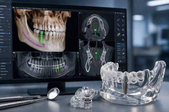
- Digital implant planning uses high-resolution 3D imaging and custom surgical guides to map the exact angle, depth, and location of an implant before surgery begins.
- Traditional freehand placement relies on two-dimensional X-rays and visual estimation during the procedure, which increases the margin for error and anatomical surprises.
- Computer-guided placement limits clinical deviation to approximately 0.5 mm, compared to up to 2.5 mm in unguided procedures.
- Shorter surgical times and smaller incisions mean less tissue trauma, significantly reduced post-operative swelling, and a faster return to daily activities.
- At D’Amico Dental Care, treatment is prosthetically driven and overseen from consultation to final restoration by Dr. D’Amico, ensuring total continuity of care.
Replacing a missing tooth is both a biological procedure and an engineering challenge. A successful dental implant must integrate firmly into living bone while supporting an artificial tooth that withstands thousands of pounds of biting pressure each day.
For decades, clinicians placed dental implants using visual judgment, manual tactile sense, and flat two-dimensional radiographs. While experienced hands often achieved good outcomes, this traditional approach left critical anatomical details to chance until the moment of surgery.
Digital implant planning changes that equation. By moving the critical clinical decisions from the operating chair to a virtual planning environment, modern implantology replaces guesswork with calculated precision.
What Exactly Is Digital Implant Planning? (And Why “3D-Guided” Is No Longer Optional in 2026)
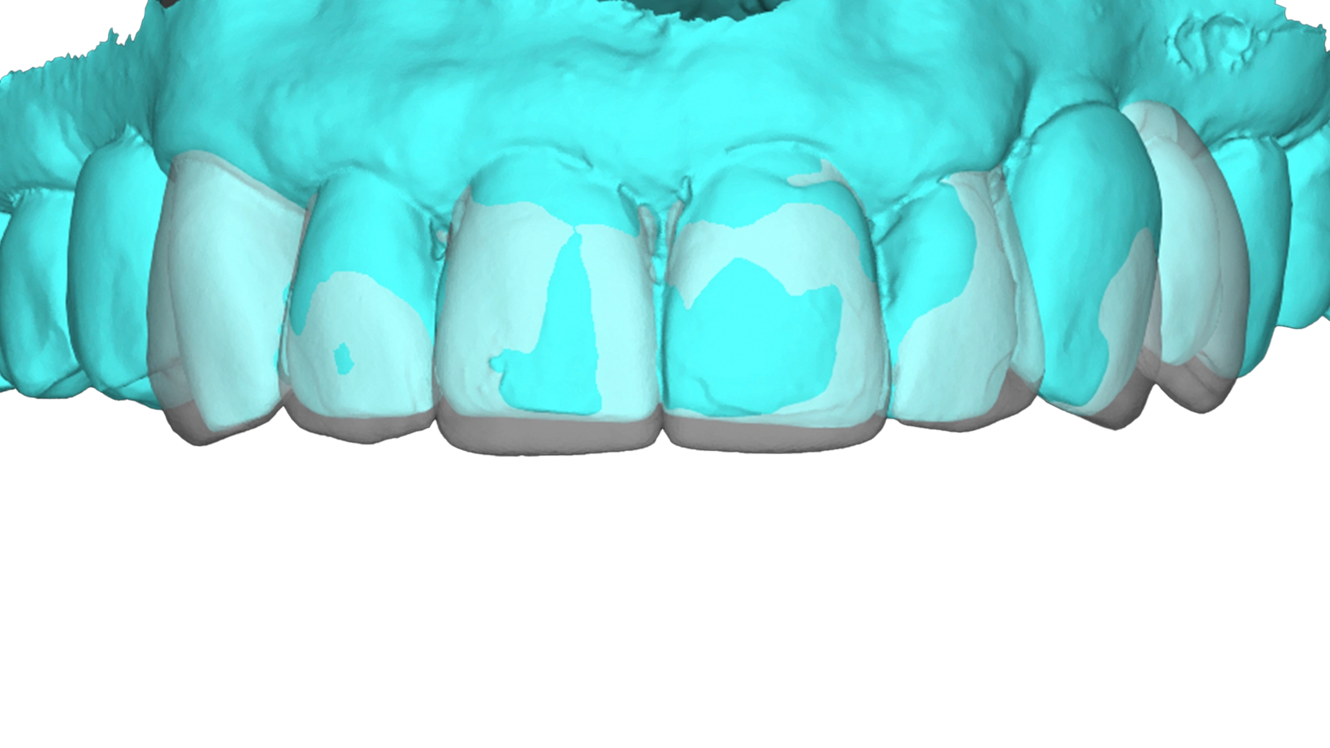
Digital implant planning is the process of building a complete, high-resolution virtual model of your oral anatomy before performing any physical procedure.
Rather than discovering the exact shape of your jawbone during surgery, your dentist plans the entire procedure inside specialized software. Every detail, including bone density, nearby nerve pathways, sinus cavities, and neighboring tooth roots, is visible in three dimensions.
By 2026, clinical standards have shifted decisively toward 3D-guided implant placement. Planning an invasive surgical procedure without volumetric diagnostics is no longer considered the benchmark of patient-centered care.
CBCT Scanning + Surgical Guides: The Technology Behind Precision Implants
The foundation of modern planning relies on two integrated technologies:
- Cone Beam Computed Tomography (CBCT): Unlike a standard dental X-ray, a CBCT scan captures a three-dimensional volumetric image of your jaw. It details bone height, bone width, and the exact position of sensitive nerve canals.
- Custom Surgical Guides: Once the optimal position is determined in the software, a bespoke acrylic guide is produced. This guide fits securely over your existing teeth or gums during surgery.
The surgical guide features precision-machined metal sleeves. These channels physically restrict the surgical drill to the pre-planned angle, depth, and position, preventing accidental deviation.
How D’Amico Dental Care ‘s Digital Workflow Differs From Traditional “Freehand” Planning
In a traditional “freehand” workflow, the dentist reviews an X-ray on a light box, makes an incision in the gum tissue, and estimates the placement angle by sight. Even skilled clinicians are limited by what the human eye can judge in a restricted surgical field.
At D’Amico Dental Care, Dr. D’Amico oversees both the surgical placement and the final restorative crown. Because our practice provides single-clinician continuity of care, your surgical plan is directly shaped by the final cosmetic goal.
We do not place an implant simply where bone happens to be easiest to reach. We evaluate your bite, smile dynamics, and bone structure together, creating a unified plan before you ever sit in the surgical chair.
Traditional Implant Planning: The 3 Hidden Risks
Traditional planning served dentistry well for many years, but it carries inherent structural limitations that modern digital tools were specifically created to solve.
2D X-Ray Limitations: Why Your Jawbone Looks Completely Different Than What the Surgeon Sees
A conventional dental X-ray takes a three-dimensional object and flattens it into a flat image. While it shows the height of your jawbone, it shows nothing about its width, thickness, or natural curvature.
Your jawbone may look dense and tall on film, yet feature sharp concavities, thin ridges, or hidden undercuts beneath the surface. With traditional methods, the clinician only discovers these anatomical variations after opening the gum tissue.
Freehand Implant Placement and the Nerve Damage Statistics You Should Know
The lower jaw houses the inferior alveolar nerve, which supplies sensation to your lower lip, chin, and teeth. Clinical studies indicate that sensory alterations from nerve disturbance occur in approximately 1% to 8% of freehand mandibular implant procedures.
Freehand placement relies heavily on visual markers and tactile resistance to avoid this vital structure. In contrast, 3D-guided surgery allows clinicians to program an intentional, non-negotiable safety buffer (typically 2 millimeters or more) directly into the surgical guide, helping protect vital nerves from injury.
The “Surprise” During Surgery That Could Have Been Predicted (And How It Affects Your Wallet)
When unexpected bone deficits appear mid-procedure during freehand surgery, the surgical plan must change on the spot. In some cases, unexpected bone grafting becomes necessary, increasing the cost and duration of your visit.
In other situations, an implant placed at an awkward angle to capture existing bone creates significant restorative challenges later. Correcting a poorly angled implant requires expensive custom abutments or compromises the final shape of your tooth, making it harder to clean and maintain over time.
Digital vs. Traditional Implant Planning: A Side-by-Side Comparison (2026 Clinical Data)
Clinical data collected across the dental profession clearly illustrates the operational differences between guided and unguided techniques.
Accuracy: 0.5mm vs. 2.5mm Deviation, What That Actually Means for Your Smile
Systematic reviews evaluate accuracy by measuring the discrepancy between where the implant was planned and where it ultimately sits in the bone.
- Digital Guided Surgery: Demonstrates an average apex deviation of roughly 0.5 mm to 0.8 mm, with minimal angular tilt.
- Traditional Freehand Placement: Commonly shows average deviations between 1.5 mm and 2.5 mm, with angular discrepancies of 7 degrees or more.
A 2 mm shift sounds minor on a ruler, but in your mouth, it is substantial. Proper spacing preserves blood supply to the delicate gum tissue between teeth, preventing artificial dark triangles from forming in your smile line.
Surgery Time: 20 Minutes vs. 90 Minutes (And Why Less Time = Less Pain)
Because all critical anatomical measurements and decisions are resolved prior to the appointment, guided implant placement often takes 15 to 20 minutes of active surgical time for a single site.
Freehand procedures often require 60 to 90 minutes. That extra time is spent verifying landmarks, taking intra-operative verification X-rays, and manually checking drill trajectories.
Shorter surgical appointments mean less time with your mouth open, less physical fatigue, and lower release of local inflammatory markers that cause post-procedure soreness.
Recovery Speed: Back to Work Tomorrow vs. Swelling for Two Weeks
Traditional freehand surgery typically requires wide incisions and extensive tissue reflection so the surgeon can directly visualize the underlying bone. This extensive tissue disruption is the primary cause of post-operative pain, bruising, and prolonged swelling.
Guided surgery is far less invasive. Because bone contours are already mapped, incisions can be kept conservative and tissue trauma is minimized. Most patients manage minor post-treatment sensations with over-the-counter pain medication and comfortably return to regular routines the following day.
How we Use Digital Implant Planning at D’Amico Dental Care
At our Wayland and Watertown offices, digital planning is not a marketing tool. It is the clinical foundation of our methodical approach to tooth replacement.
Planning Starts With the Final Restoration
We practice prosthetically driven implant dentistry. This means we design the final crown first, evaluating proper bite mechanics, harmonious tooth proportions, and natural aesthetic contours.
Once the ideal position of the visible tooth is confirmed, we plan the titanium foundation underneath it. The foundation supports the crown, not the other way around.
Bone, Soft Tissue and Anatomy Are Assessed in Three Dimensions
During your diagnostic exam, Dr. D’Amico analyzes your bone volume, gum contours, and adjacent anatomical structures using advanced 3D scanning.
If site-preparatory steps like bone grafting are necessary, they are identified and discussed calmly before treatment begins. Nothing is left to mid-procedure improvisation.
The Implant Position Is Planned Around the Whole Treatment
Because Dr. D’Amico manages your entire treatment arc, our planning accounts for the full healing journey:
- Protecting long-term bone-to-implant stability.
- Designing appropriate contours for soft-tissue healing.
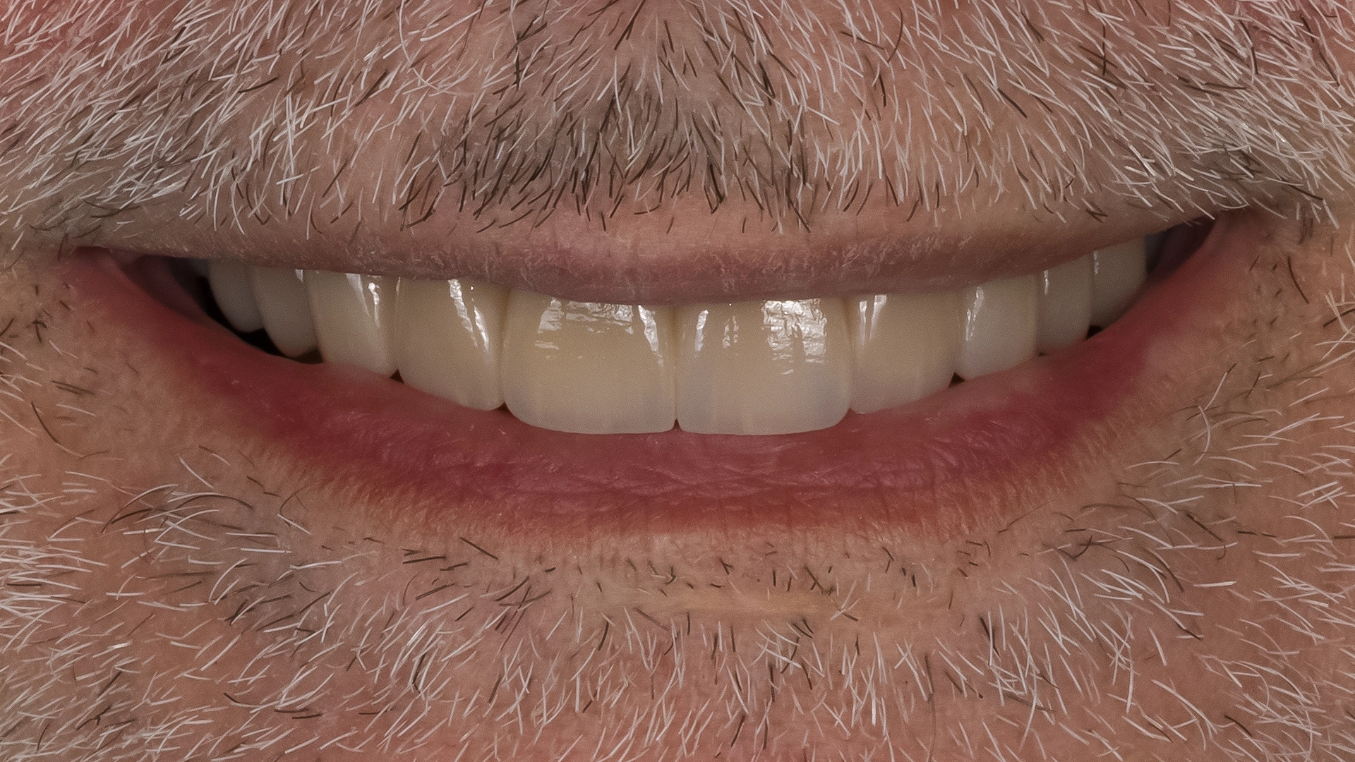
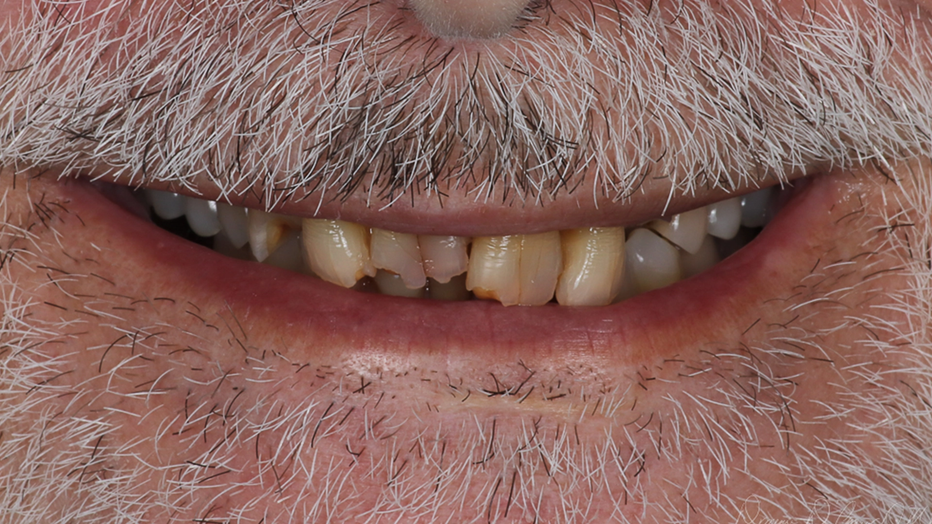
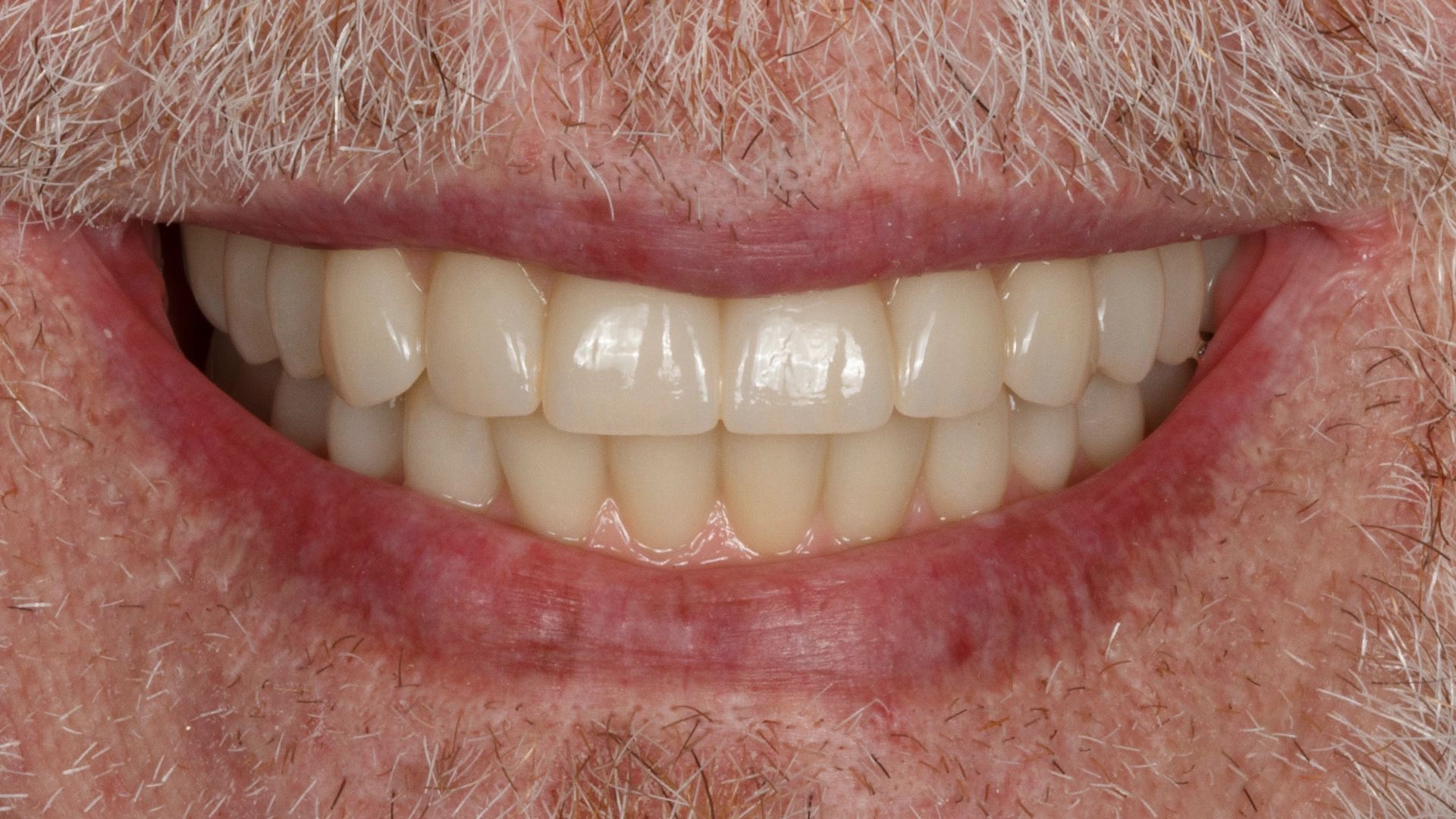
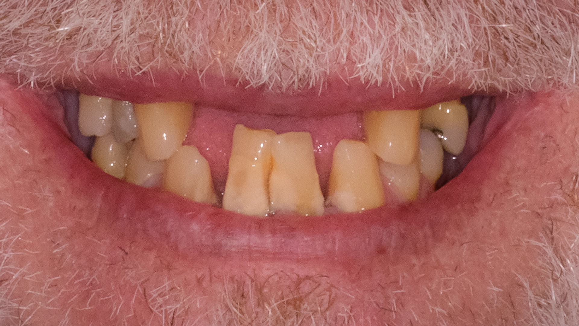
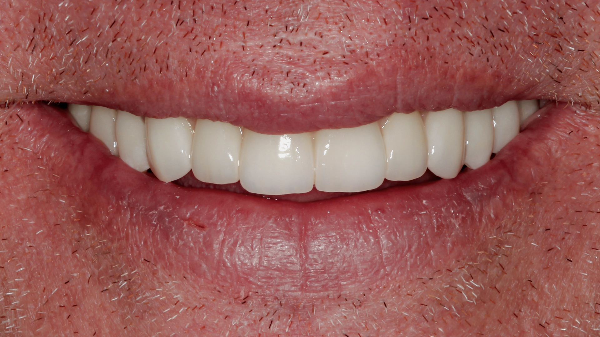
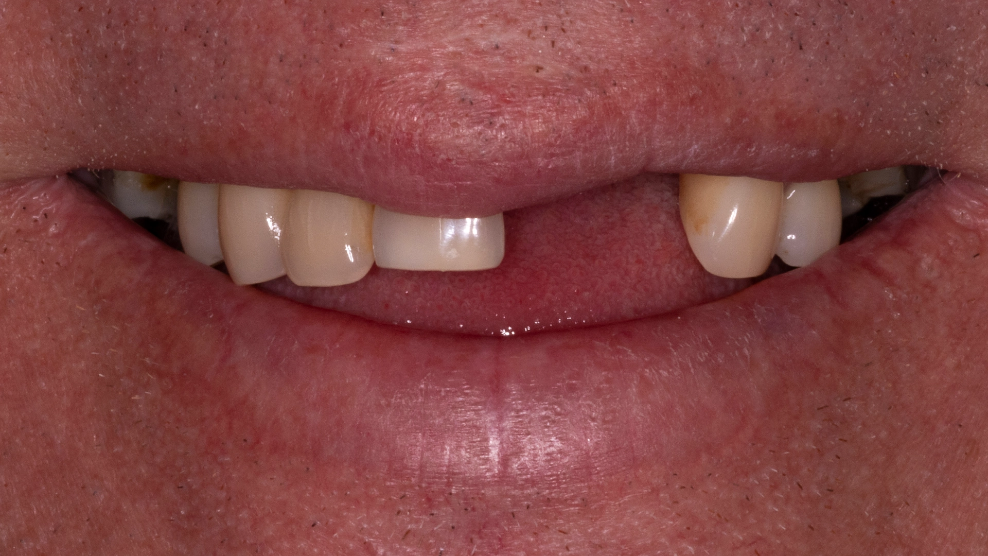
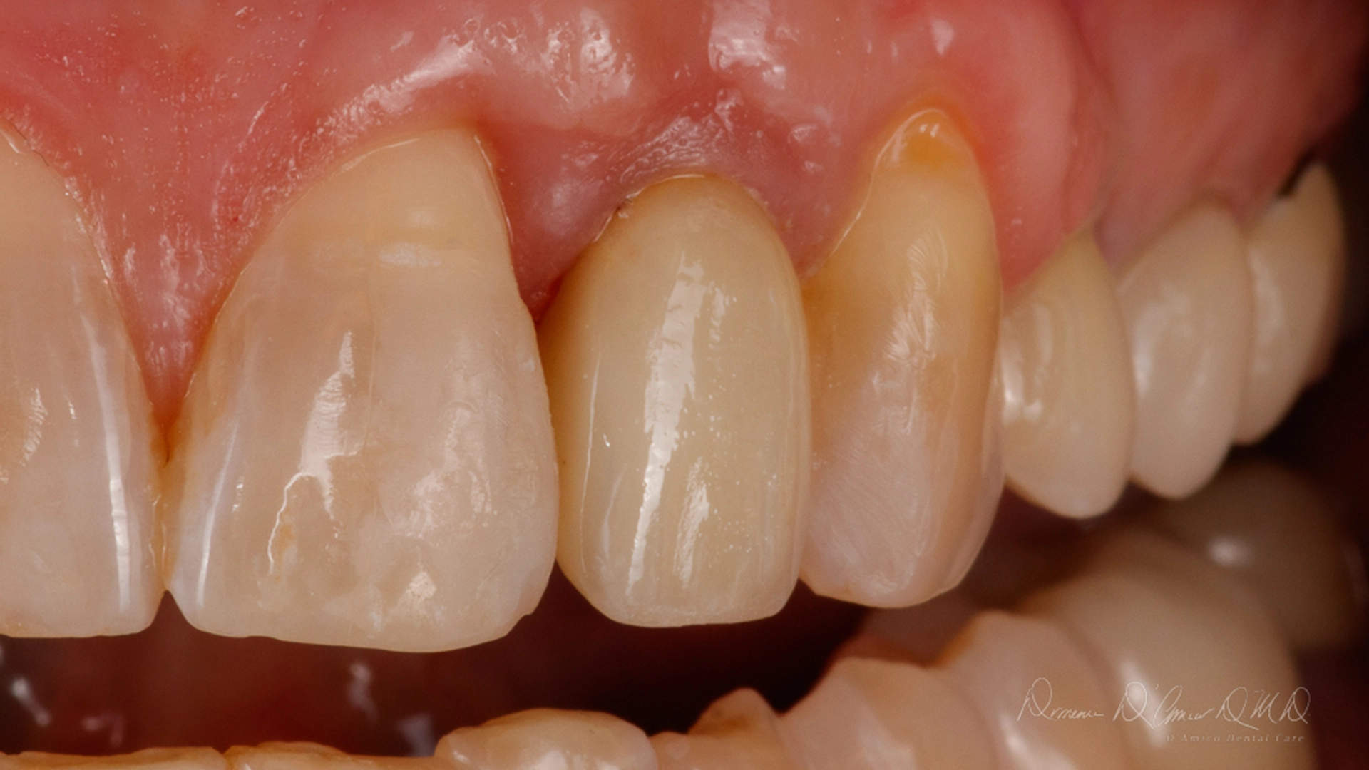
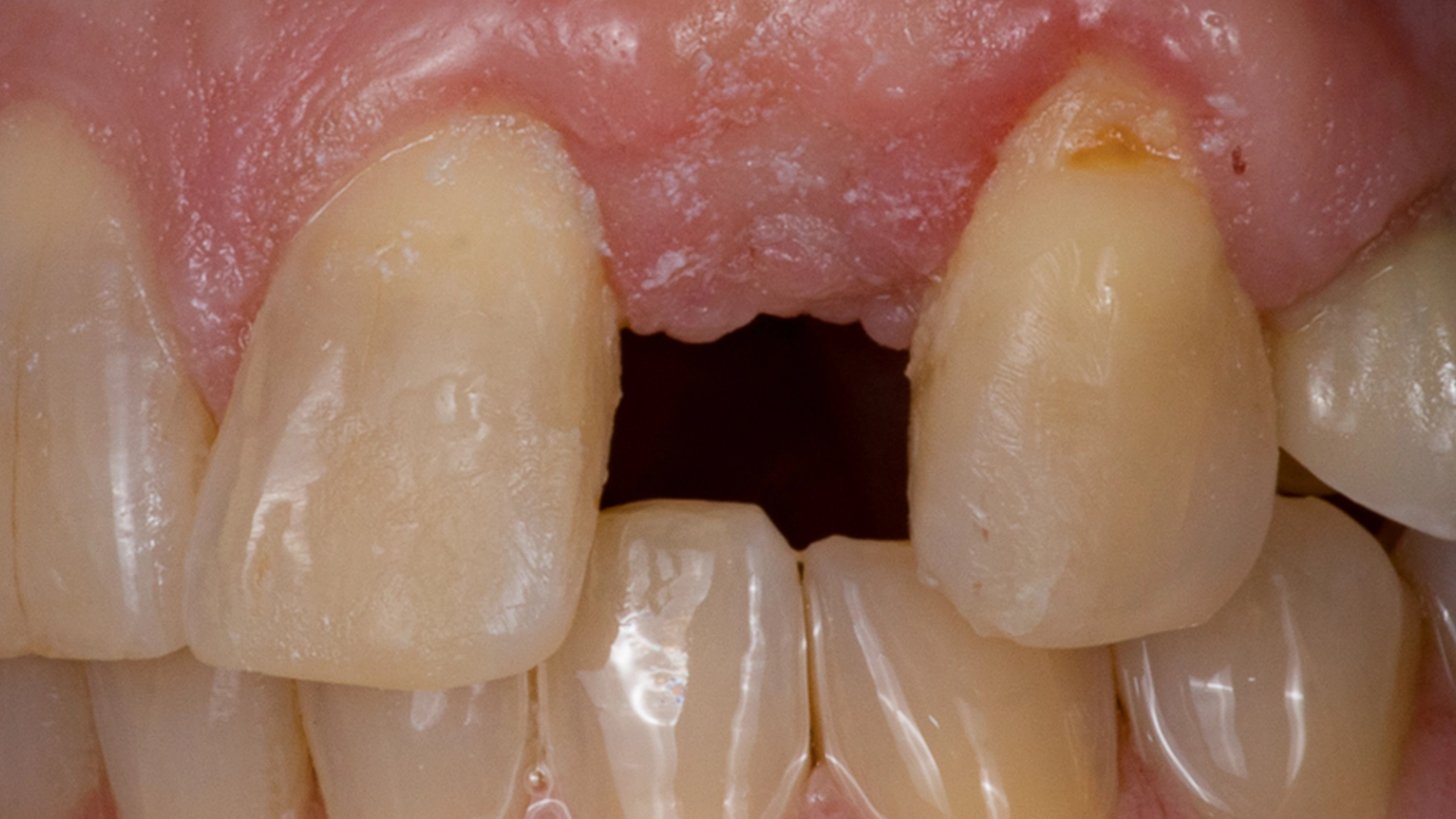
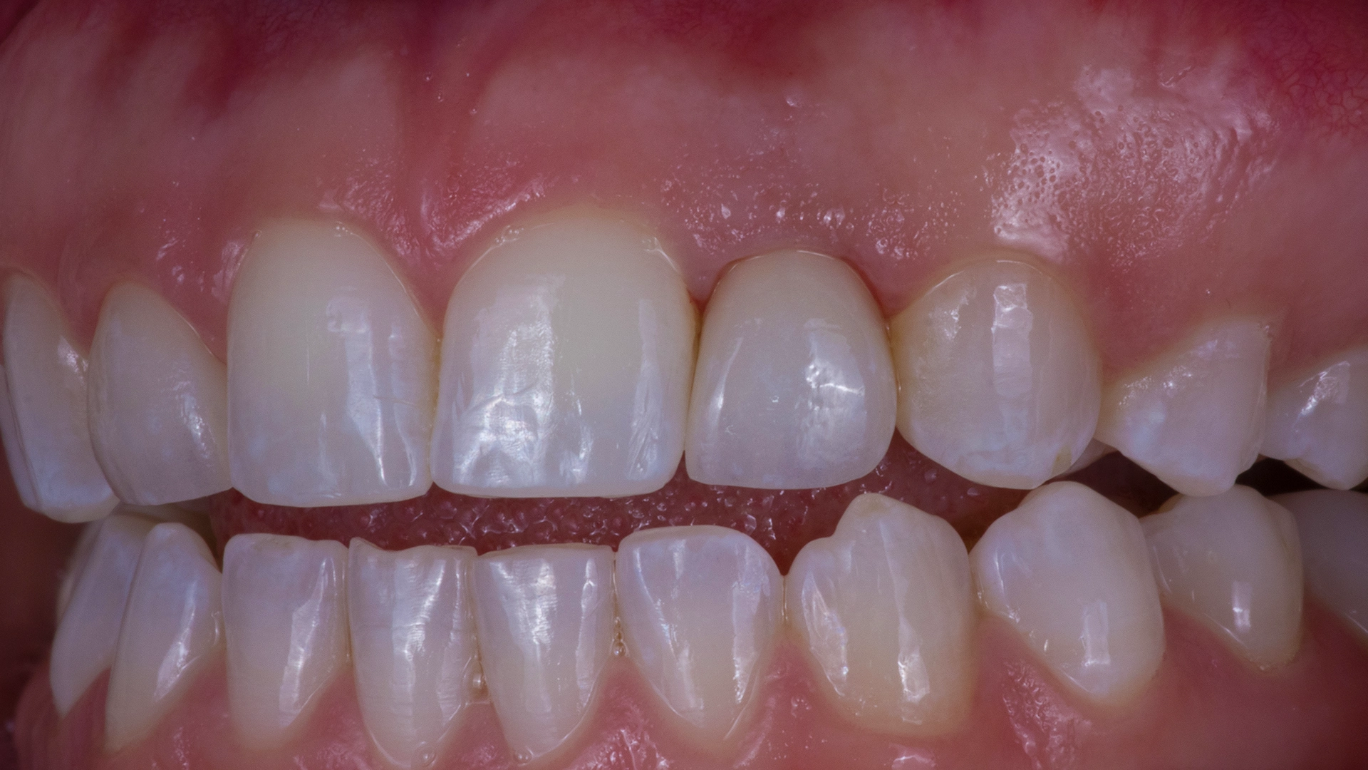
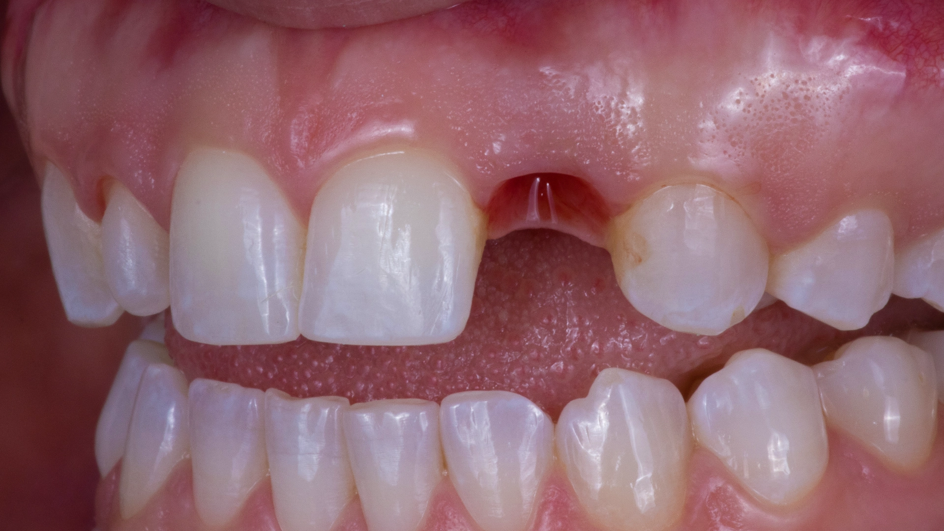
- Preparing for a provisional (temporary) restoration when appropriate, ensuring you do not have to leave our office with a visible gap while your implant integrates.
What Should You Do If You Are Considering Dental Implant Treatment
Deciding to restore your smile with dental implants is an important health decision. You deserve clear, detailed explanations rather than hurried recommendations.
Start With an Assessment of Your Individual Anatomy
The most reliable first step is a comprehensive diagnostic consultation. A proper clinical evaluation examines your unique bone health, overall medical history, and aesthetic goals to determine whether an implant is your most dependable long-term option.
Take the Next Step Toward a Well-Planned Smile
If you are exploring tooth replacement options in the Wayland or Watertown areas, we invite you to experience the clarity of a carefully planned process.
Contact D’Amico Dental Care to schedule your initial diagnostic consultation. We will evaluate your anatomy, explain every stage of care, and provide a clear, structured treatment plan designed for long-term health and stability.
Frequently Asked Questions
1. What is the main difference between 3D-guided digital surgery and traditional freehand implant placement?
Direct Answer: The primary difference is the complete removal of intra-operative guesswork through pre-surgical computer simulation.
In traditional implantology, the clinician evaluates flat 2D X-rays, reflects the gum tissue, and estimates the drill trajectory and depth visually and by tactile feel during surgery. In contrast, digital planning reconstructs the jawbone and surrounding anatomy in dedicated 3D software prior to treatment. A custom surgical guide is then fabricated and secured in the mouth, physically locking the surgical drill to the pre-planned angle, depth, and sub-millimeter coordinates.
2. Does digital guided implant placement involve less pain, swelling, and bleeding?
Direct Answer: Yes; digital implant placement offers significantly faster recovery due to minimal soft-tissue trauma.
In unguided procedures, clinicians typically make extensive incisions and elevate large gum flaps to directly visualize the underlying bone ridge, which is the primary cause of post-operative pain, bruising, and prolonged swelling. With 3D digital planning, bone boundaries are fully known in advance. This allows for conservative, tissue-sparing incisions and faster surgical times, enabling most patients to return to routine daily activities the following day with minimal discomfort manageable by over-the-counter pain medication.
3. If I have bone loss in my jaw, can I still receive dental implants with this method?
Direct Answer: Yes; 3D digital planning maximizes the use of existing bone and identifies any bone grafting requirements well before surgery.
Reduced bone volume is rarely an insurmountable barrier. Virtual planning tools allow the clinician to assess bone width, height, and density in volumetric cross-sections. This enables the doctor to identify areas of solid cortical bone and optimize implant angulation to maximize available volume. If preparatory procedures such as bone grafting or sinus augmentation are necessary, they are sequenced transparently in advance rather than discovered unexpectedly mid-procedure.
4. What does “prosthetically driven” implant planning mean, and how does it affect aesthetics?
Direct Answer: Prosthetically driven planning designs the final visible crown first, placing the implant foundation where it best supports a natural-looking tooth and balanced bite.
In traditional workflows, implants were often inserted wherever bone was easiest to access, occasionally forcing the restorative lab to craft angled or bulky artificial teeth that were hard to clean and visually unnatural. With a prosthetically driven approach, your final smile contour, tooth proportions, and bite dynamics are designed virtually first. The implant post is then placed underneath at the ideal angle to replicate how a natural tooth emerges from the gum line.
5. What risks associated with traditional freehand placement are prevented by digital guidance?
Direct Answer: Digital guidance helps prevent angular misalignment, nerve injury, and damage to adjacent tooth roots.
In the lower jaw, the inferior alveolar nerve supplies sensation to the lower lip and chin; unguided freehand drilling in compromised bone heights carries a higher risk of nerve disturbance. Digital planning incorporates a verified safety buffer directly into the guide to keep instruments safely away from critical nerve pathways. The physical guide also prevents the drill from inadvertently drifting toward healthy adjacent roots or piercing through cortical bone plates.
6. Will I have a gap in my smile while the implant integrates with the bone?
Direct Answer: No; customized provisional restorations are planned ahead of time so you do not have to live with a visible gap.
Preserving your appearance and confidence throughout treatment is a standard clinical priority, especially in the aesthetic smile line. Because diagnostics and dimensions are captured before the appointment, a provisional (temporary) tooth can be fabricated in advance. When primary stability and clinical assessment permit, this temporary restoration is secured immediately after placement, keeping your smile intact while osseointegration occurs over the subsequent months.
7. Where is D’Amico Dental Care located, and which surrounding communities do you serve?
Direct Answer: We operate two modern dental practices in Wayland and Watertown, proudly treating patients across the Greater Boston and MetroWest regions.
Beyond our local communities in Wayland and Watertown, a substantial portion of our patient base travels from nearby towns, including Newton, Wellesley, Weston, Sudbury, Belmont, Waltham, and Cambridge. Patients seek out D’Amico Dental Care specifically for our predictable digital workflows and the clinical continuity provided by Dr. D’Amico, who oversees the entire implant pathway from diagnostic planning to final crown restoration.
8. Is digital implant planning cost-effective compared to traditional placement?
Direct Answer: Yes; the precision of guided placement helps prevent costly complications, surgical revisions, and expensive corrective components.
While 3D imaging and custom surgical guide manufacturing involve an initial diagnostic investment, they significantly reduce the risk of implant failure, biological complications, and the need for expensive custom angled abutments to correct misaligned implants. By ensuring that the foundation is positioned correctly the first time, digital planning provides a predictable, long-term solution that protects your health and your financial investment.